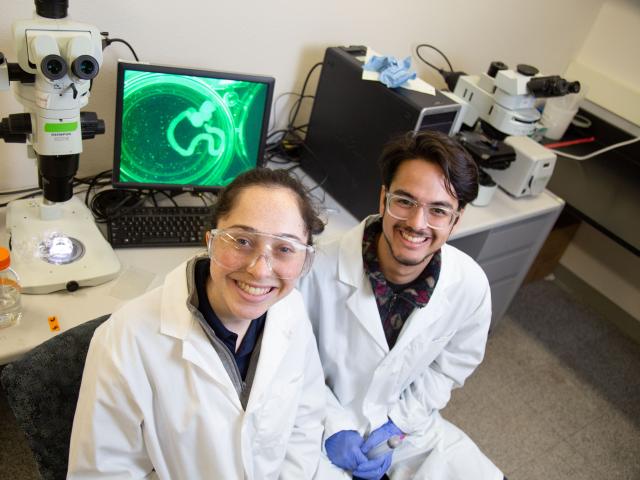
Distinguished Professor Walter Leal, recently held a second online symposium on COVID-19. The first and second symposiums were attended by several thousand viewers and covered a range of topics related to COVID-19.
The rapamycin-sensitive TOR kinase, an essential regulator of cell growth, assembles into two multi-protein complexes, termed TORC1 and TORC2. Taking advantage of two homologous TOR kinases in budding yeast, a minimal assembly specificity (MAS) domain was identified that governs their assembly into TORC1 versus TORC2. Based on a recent structural study of TORC2 by Loewith and colleagues, the TORC2 MAS domain (depicted in blue) is predicted to be involved in quaternary interactions (shown in yellow, green, and red) important for TORC2 specificity and assembly.
Confocal micrograph of an 11 day post fertilization zebrafish gonad showing germ cells (blue) surrounded by epithelial layer of somatic gonad cells expressing the fibroblast growth factor (Fgf) ligand fgf24 (green) and an inner mesenchymal layer expressing the Fgf-responsive gene etv4 (red). Photo credit: Dr. Dena Leerberg. For more information, see Leerberg, D.M., Sano, K. and Draper, B.W. (2017) Fibroblast growth factor signaling is required for early somatic gonad development in zebrafish. PLoS Genetics, 13(9): e1006993.
Zebrafish early-stage oocytes (red) are surrounded by cyp19a1a-expressing theca cells (green; all nuclei are stained blue). cyp19a1a encodes the aromatase enzyme that produces estrogen and is required for female sex determination. In female zebrafish, oocytes produce the Bmp15 signaling ligand, and while bmp15 mutants can become female, they fail to maintain female sex differentiation and sex-revert to fertile males during the juvenile stage of development, in part because they fail to maintain expression of cyp19a1a. See more at http://dx.doi.org/10.1371/journal.pgen.1006323
LET-99 is a G-protein regulator that localizes to the presumptive site of cleavage. YFP::LET-99 localization through cytokinesis of a one-cell C.elegans embryo. The image has been pseudo-colored to show the range of YFP::LET-99. Brighter represents greater protein localization.
The crystal structure of the Kinesin-5 BASS domain tetramer: (A) Side view of the crystal structure of the KLP61F BASS tetramer (residues 640–796 shown) colored in rainbow, starting with N-termini in blue traversing to C-termini in red, respectively. Four monomers pack as anti-parallel pairs of anti-parallel coiled-coil dimers. (B) shows the BASS structure rotated 90° around the filament axis relative to panel A. The dimensions of the BASS tetramer bundle structure are shown. (C) Side view of the BASS tetramer, with two BASS anti-parallel dimers colored in blue and red, respectively. (D) Detailed interaction between two monomers in the BASS anti-parallel dimer. (E) A 60° rotated view of D. - See more at: http://elifesciences.org/content/3/e02217#sthash.9VMjrUdO.dpuf
C. elegans male germ cells: Top, gonad dissected out of worm stained with RAD51 recombinase (green), chromatin mark, H3K9me2 (red) to mark the X chromosome, and DAPI (DNA, blue) . Bottom, individual cells from different germline stages: left, mitosis (tubulin, green; CENPA, red; DAPI, blue); middle, meiotic prophase (pachytene): top, aligned homologous chromosomes (synaptonemal complex, red and green; DAPI, blue); bottom, nuclear pore complex (yellow) and DAPI (blue) and right, the meiosis I division (tubulin, green; membrane protein, red; DAPI, blue).
Proposed model for kDNA organization in Trypanosomes
Superresolution (SIM) imaging of mammalian cells defective for mitochondrial division expressing mitochondrial-targeted DsRed (mito-DsRED) and ER-targeted GFP (GFP-HDEL.
Electron tomography of integrin configuration on the limiting membrane of SAOS-α2β1 cells. The missing wedge effect of ET has been resolved to achieve the isotropic resolution in all dimensions of the structures. This allows the tracking of the distribution of α2-integrin in an endosome intracellularly.
Top, a revised paradigm for the tubulin cofactors and the Arl2 GTPase as a chaperone involved in the biogenesis and degradation of αβ-tubulin dimers. Bottom, The effect of tubulin cofactors/Arl2 chaperones on soluble pool of αβ-tubulin in the cytoplasm on the dynamic microtubule polymerization.
Image of kinetoplast DNA (kDNA), mitochondrial DNA found in trypanosomes, produced by Lara Ibrahim, who is now a PhD student at the Scripps Institute in San Diego. Lara was assisted by Samantha Lewis (postdoc in the Nunnari lab) and from Fei Guo.
News and Events

After 23 years, the Life Sciences building is getting a new name that befits its purpose: Melvin M. and Kathleen C. Green Hall.
News and Events

MCB is proud to announce that Dr. Dan Starr and Dr. Gant Luxton have been selected as a Allen Distinguished Investigators, by the Paul G. Allen Frontiers Group.
News and Events

Dr. Oliver Fiehn has been named in the annual Highly Cited Researchers 2020 list released by the Web of Science Group, which compiles statistics on scientific publishing. The list identifies scientists and social scientists who have published multiple papers ranking in the top 1 percent by citations in a particular field and year, over a 10-year period.