
Worms Reveal Just How Cramped Cells Really Are
UC Davis Research Shows Cytoplasm is less like a Pool than a Crowded Concert Venue
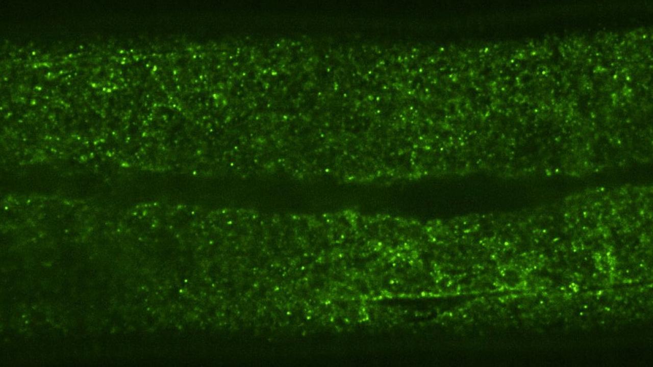
In a new study published in Science Advances on September 10, a team of UC Davis researchers tracked the movement of fluorescent particles inside the cells of microscopic worms, providing unprecedented insights into cellular crowding in a multicellular animal. They found that the cytoplasm inside the worms was significantly more crowded and compartmentalized than in single-celled yeast or mammalian tissue culture cells, which are more commonly used to gauge internal cellular dynamics.
This difference highlights the importance of studying cellular processes in living animals rather than cell culture, says first author Xiangyi Ding, a Ph.D. candidate in the Integrative Genetics and Genomics graduate group.
“This changes everything. Crowding in a cell affects any process that depends on molecule movement and interaction, including drug delivery, disease progression, and how cells respond to stress.”